-
Phone
+385 91 444 45 11 -
Email
info@poliklinika-skara-kolega.hr
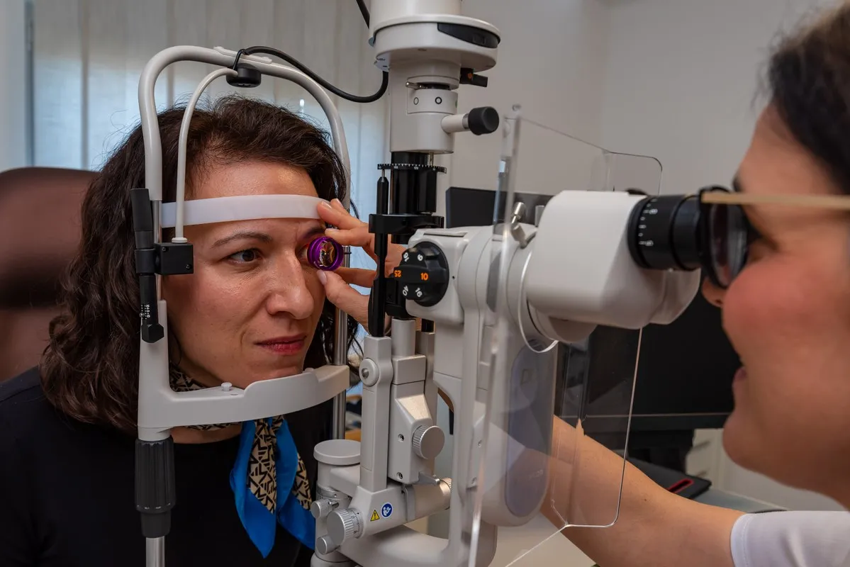
The specialist ophthalmological examination includes visual acuity examination, determination of prescriptions for glasses, measurement of intraocular pressure, and examination of the anterior and posterior eye segments (fundus).
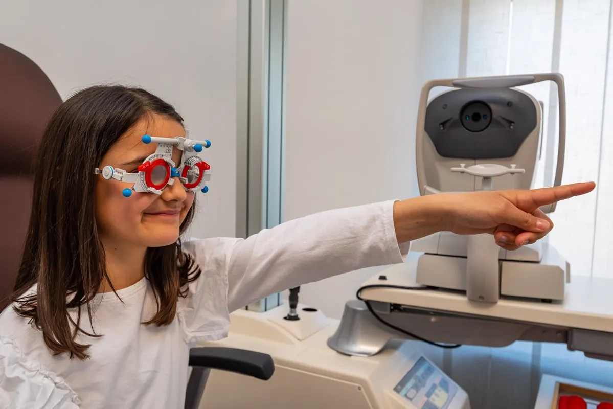
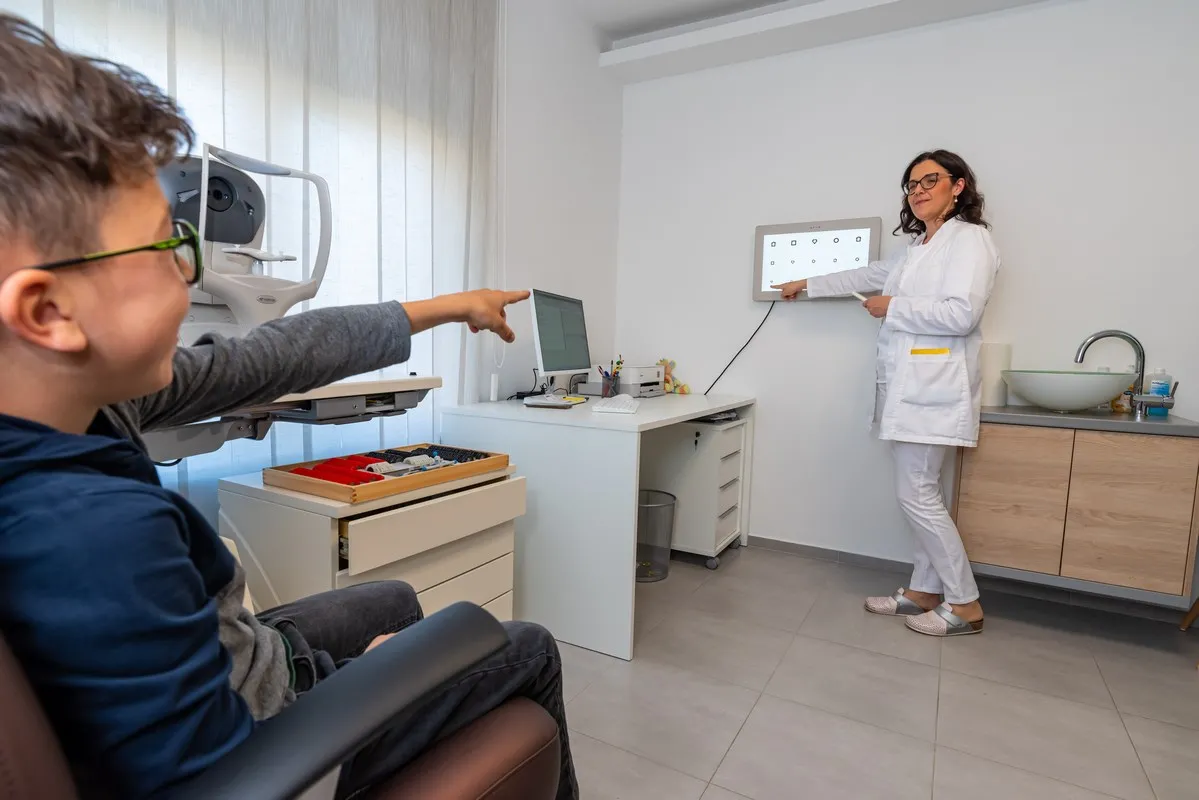
The pediatric ophthalmic examination includes a comprehensive examination of the eyes and vision in children from birth to adulthood, including prematurely born children (premature infants). The examination involves vision assessment (visual acuity), stereo vision, examination of the anterior eye segment, and fundus examination using an indirect binocular ophthalmoscope, as well as measuring intraocular pressure. Through the examination, we can determine whether your child has refractive errors and needs glasses, whether the child has amblyopia (lazy eye), and whether there is strabismus (eye misalignment), eye wandering, or squinting. A complete ophthalmic examination for a child is never too early because if there is a problem, the best treatment results are achieved with an early start of treatment. It is recommended that a child undergo a thorough ophthalmic examination in the first 4 years of life. If you notice any vision problems, eye wandering, or other issues, the examination should not be delayed but conducted as soon as possible.
Strabismus is a disorder of eye position or mobility where the eyes do not have the same direction of gaze. It most commonly occurs in childhood but can also occur in adulthood. It can be congenital or acquired after birth. It arises due to vision disorders (refractive errors, nystagmus, amblyopia), eye diseases (tumors, albinism, retinopathy of prematurity), injuries, neurological conditions (stroke, myasthenia gravis, Parkinson's disease, etc.), internal diseases (diabetes), and endocrine diseases (thyroid diseases).
The examination includes assessing the degree of stereo vision, eye mobility, measuring the eye deviation angle, and the prism adaptation test. Skiascopy is often necessary to determine the objective eye prescription.
With a complete ophthalmic examination, glaucoma treatment includes OCT (optical coherence tomography), visual field testing, pachymetry, and gonioscopy. All services can be done at our clinic without waiting and in a single visit.
Retinal diseases include senile macular degeneration, diabetic retinopathy, macular holes, epiretinal membranes, retinal ruptures and detachments, retinal dystrophies, and retinal nevi. Alongside modern diagnostics, we apply treatment with laser and intraocular injections.
The cataract examination includes a comprehensive examination with pupil dilation and measurement of intraocular pressure. In preparation for surgery, an extended examination of the fundus of the eye, OCT of the macula, and optic nerve are performed. The calculation of the intraocular lens is done using a new and modern non-contact optical biometer, which provides an accurate calculation of the intraocular lens that will be implanted in place of the cloudy natural lens. During the examination, it is important to determine through conversation about desires, habits, professions, and hobbies which intraocular lens is the best choice for each patient.
Monofocal intraocular lenses have a single focus, providing good distance vision without glasses. For near vision, glasses are required. It is also possible to achieve good near vision without glasses, depending on the patient's preferences, with the need for glasses for distance vision.
Toric monofocal lenses are suitable for patients with astigmatism, providing good distance vision, while reading glasses are needed.
Monofocal lenses with extended focus provide good distance and intermediate vision up to 50-60cm. Glasses will be needed for reading at a closer distance.
Multifocal lenses have multiple focal points, allowing good vision at various distances. Bifocal lenses have two focal points, providing good vision for both distance and near, but intermediate distances such as working on a computer may remain unclear, requiring glasses. Newer generations include trifocal lenses, offering clear vision at all three distances: distance, near, and intermediate. With these lenses, activities like reading, writing, using a computer, watching TV, driving, and engaging in sports can be done without glasses.
Contact lens examination includes checking visual acuity, measuring corneal curvature - keratometry, examination of the anterior eye segment, application of a trial contact lens, and education on independent insertion and removal of contact lenses.
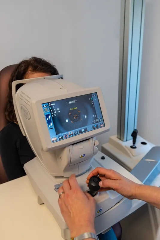
Visual acuity examination includes assessing visual acuity without glasses (natural vision), determining the prescription with an automatic computerized refractometer, and correcting and determination of the prescription for near and far-vision glasses.
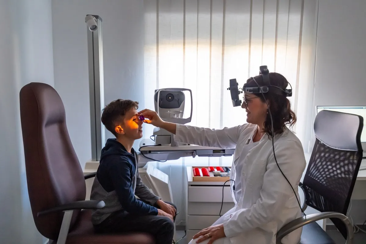
An objective method for determining the actual eye prescription. Eye drops to dilate the pupils are instilled 2 or 3 times every 15 minutes, and then, in a dark room, the prescription is determined using light from a skiascope. During this process, the child sits either on a chair or in the lap of a parent. This method of determining the eye prescription does not require the child's knowledge or communication skills. The child does not need to speak or read. After determining the prescription, glasses are prescribed as needed.
Determining the position and mobility of the eye, and coordination between both eyes. The mobility of each eye separately (ductions) and both eyes together (versions and vergences) is assessed. If there is strabismus, squinting, or eye deviation, the deviation angle is measured in all directions of gaze, both at near and far distances. It is measured with prisms, and the value is expressed in prism diopters (PD) or degrees (°). Orthoptic status needs to be determined in children, strabismus, and double vision.
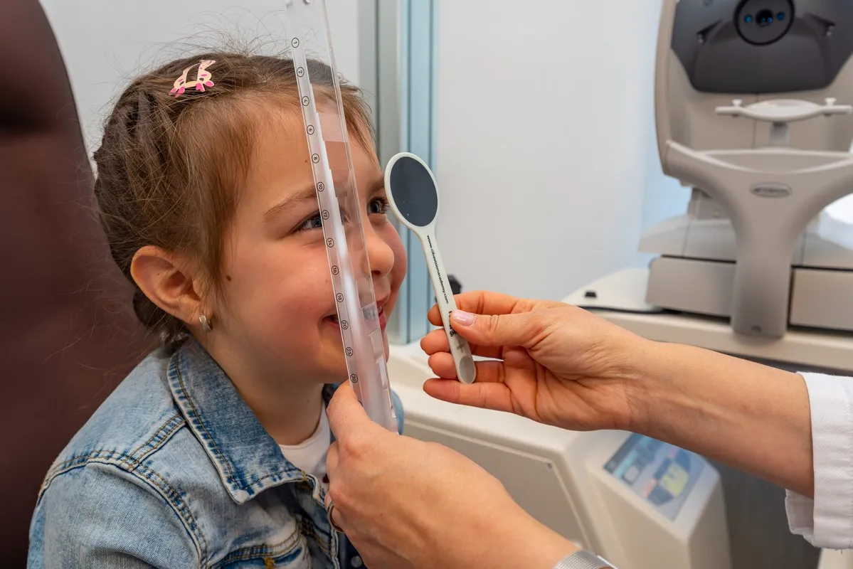
Determining the degree of stereo vision, which is an important indicator of binocularity and coordination between both eyes. It is determined using the Bagolini test, Titmus test, and Lang test.
Determination of the prescription on the device, computerized autorefractometer.
Ophthalmic examination measuring corneal curvature, is one of the most important factors for determining contact lens fitting. It is measured using a computerized keratometry device
We examine the front segment of the eye (eyelids, conjunctiva, sclera, cornea, anterior chamber, iris, and lens) using a slit lamp microscope.
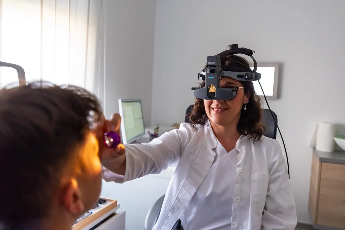
After instilling eye drops to dilate the pupils and waiting for about 15 minutes for the pupils to expand, the lens and the back of the eye, the fundus (vitreous, optic nerve, macula, retina), are examined with the help of a magnifier. The examination can be performed on a slit lamp or with an indirect binocular ophthalmoscope used for examining children and patients with spine problems who are unable to assume an adequate position for examination on a slit lamp.
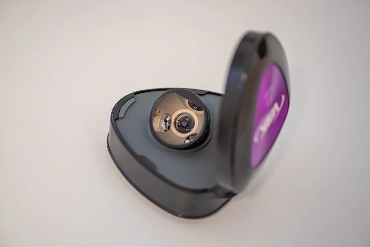
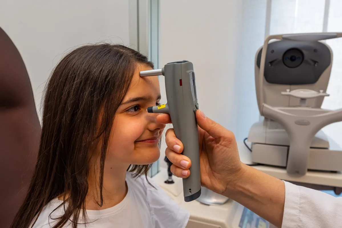
It is measured with an applanation tonometer or a handheld non-contact tonometer. Applanation tonometry is a contact method performed on a slit lamp in a seated position after instilling local anesthetic eye drops. The handheld tonometer is a portable device used for minimal contact measurement of intraocular pressure. It does not require instillation of local anesthetic and can be measured in all body positions.
Examination of the anterior chamber angle, the anatomical angle between the cornea and iris, using a special gonio-lens, a mirrored prism. It is crucial for determining the type and method of glaucoma treatment. The examination is performed by placing a mirrored prism on the corneal surface, and the examination is not painful because local anesthetic drops are instilled beforehand. To display the anatomical structure of the chamber angle in our clinic, we also use OCT examination of the anterior eye segment, which does not come into contact with the eye.
Tear Film Fluorescein Dye Test using fluorescein strips, which we use for corneal damage diagnosis and dry eye diagnosis.
Test to determine the amount of tears in dry eye disease.
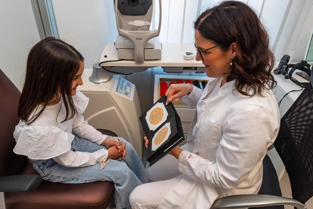
Color vision tests are used to determine complete or partial color vision impairment (color blindness). We use Ishihara plates.
© Copyright 2026 Poliklinika Škara Kolega. All rights reserved. Made by ASPEKT