-
Phone
+385 91 444 45 11 -
Email
info@poliklinika-skara-kolega.hr
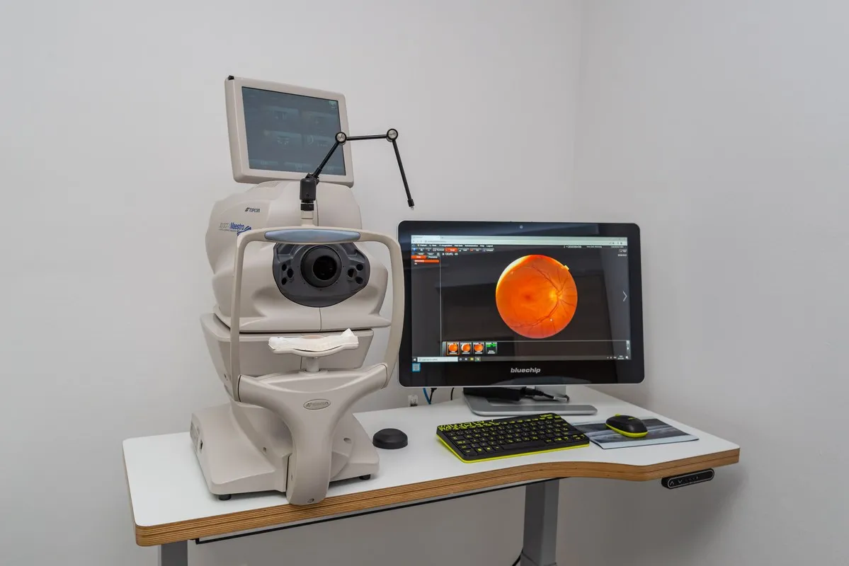
We photograph your eye and present you with an image of the eye as seen by an ophthalmologist, whether it's the anterior segment or the posterior eye. It is advisable to monitor individual changes in the eye that change over time, such as nevi (moles) on the eye.
Measuring corneal thickness is essential for the correct interpretation of intraocular pressure measurement results in cases of suspected or diagnosed glaucoma. It is measured using a contact ultrasound pachymeter or, non-contact, with the help of an OCT device.
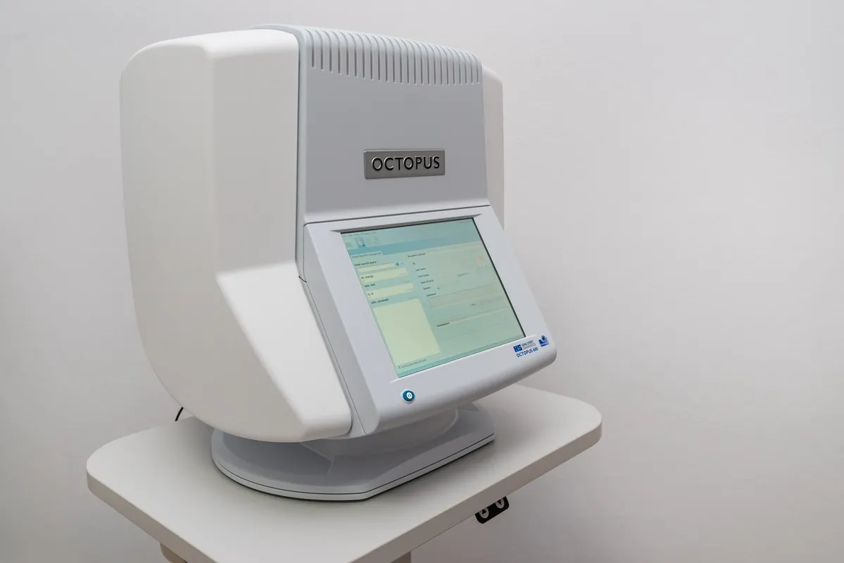
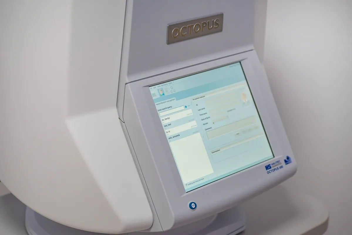
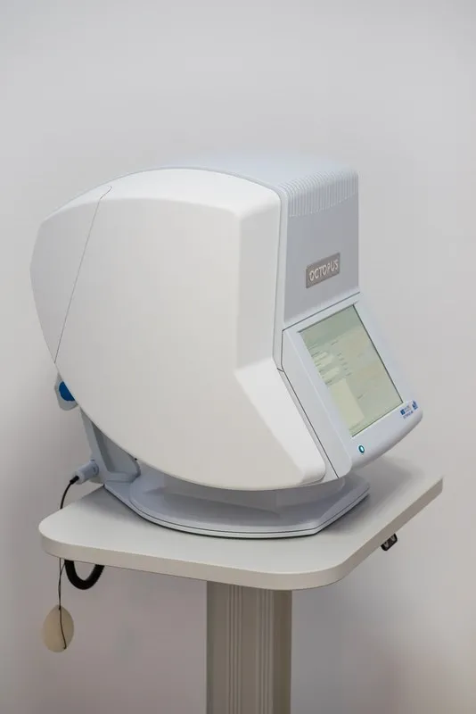
The visual field or automated perimetry is the most important functional examination. It determines the width we see without turning our heads. It is extremely important in glaucoma and many neurological diseases. The Octopus 600 visual field allows various programs for targeted detection and monitoring of disease progression in all its phases. It is highly sensitive and avoids errors caused by loss of focus or patient fatigue. The examination takes from 15 to 30 minutes, and the result is obtained in both visual and written form.
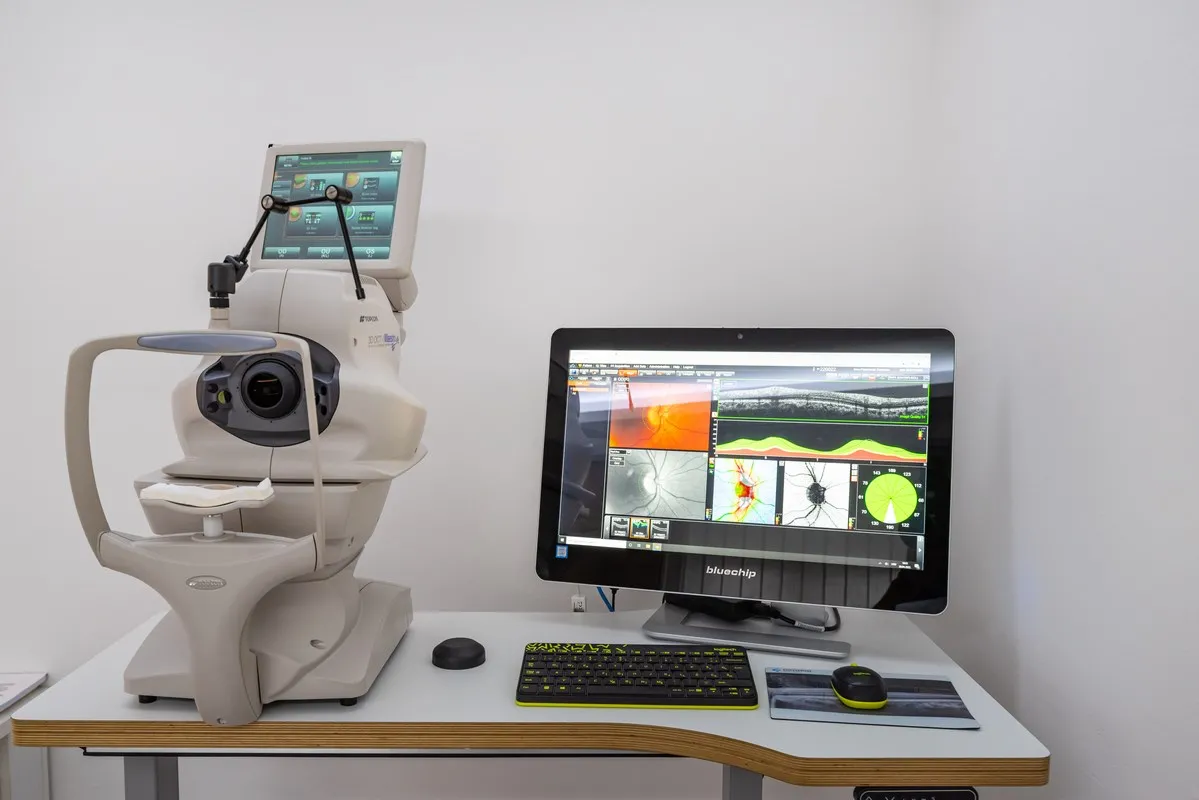
Optical coherence tomography is a method of imaging the eye at a microscopic level using laser technology. The high resolution, exceptional speed, and accuracy of our 3D-OCT device enable imaging of the cornea, anterior chamber angle, optic nerve, macula, and retina. It is extremely important for diagnosing and monitoring diseases such as glaucoma, age-related macular degeneration, diabetic retinopathy, macular ruptures and edema, epiretinal membranes, retinal detachments, corneal and anterior chamber angle diseases, and corneal thickness measurement (pachymetry). All findings are photo-documented, and the patient receives both visual and written reports.
A simple and painless examination of the back of the eye and orbit. It is used in people with blurred optical media, such as advanced cataracts, corneal opacities, bleeding inside the eye (hemophthalmus), retinal detachment, or intraocular tumors. It is performed by gently applying an ultrasound probe to closed eyelids, which are pre-gelled.
Biometry is a non-contact method of measuring the length of the eyeball, the curvature of the cornea, and the thickness of the eye lens, based on which the power of the lens implanted during cataract surgery is calculated. It is also used in monitoring the progression of myopia (nearsightedness). The scanning takes a few minutes, and no preparation is required. The calculation result is obtained immediately after the examination.
© Copyright 2026 Poliklinika Škara Kolega. All rights reserved. Made by ASPEKT